Vertebral Body Height Measurement
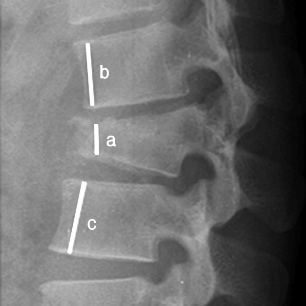
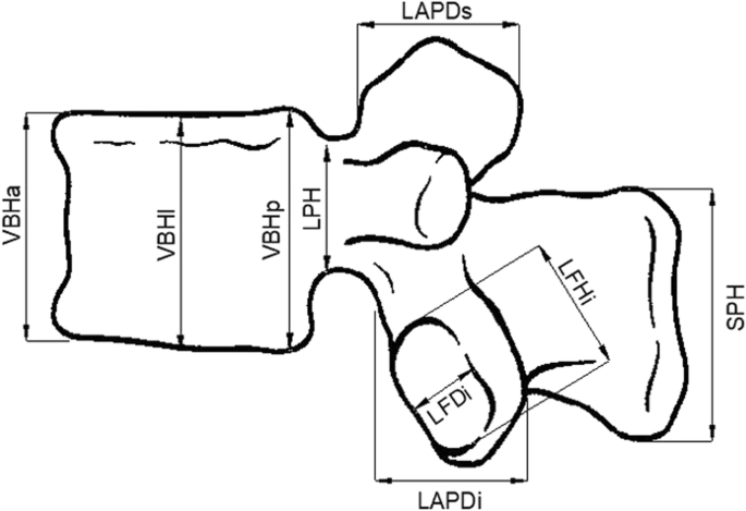
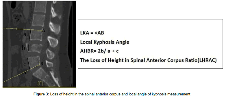
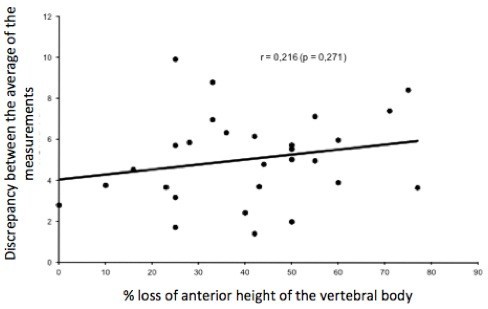
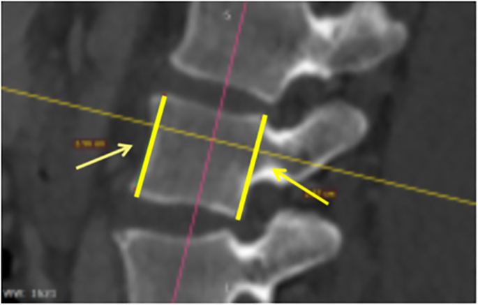
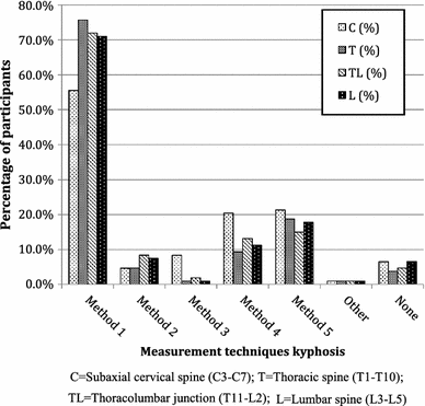
Previously researchers used different measurement methods to assess the degree of vertebral body collapse by using posterior wall height as the reference vertebral body compression ratio vbcr or the percentage of anterior height compression pahc which uses the mean height of segments adjacent to a healthy vertebral body as the reference value 8 11. The objective of this study was to analyze the spinal dimensions of the ais spine in 3 d in vivo by measurement of the total length of the spine and height width depth ratios of the vertebral. Criteria for deformity should allow for this variation to avoid misdiagnosis. Normal vertebral dimensions were found to vary from vertebra to vertebra. The lvf group had significantly lower dorsal and ventral t12v body heights both p0001. This study used previous measurement methods to assess the degree of vbhl and ka compare and examine differences between various measurement methods and examine the correlation between relevant measurement parameters and intravertebral cleft ivc in the vertebral body.
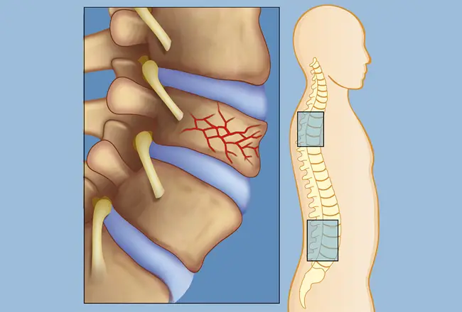
The normal lower bound for relative change in measured vertebral heights ranged between 10 and 16 anteriorly and 11 and 19 posteriorly. The average dorsal t12v body height was 2125164 mm in the control group and 2011149 mm in the lvf group. Anterior vertebral height remained the same from the third to the fifth vertebra but the posterior vertebral height decreased. For compression fracture vertebral body height loss vbhl and kyphotic angle ka are two important imaging parameters for determining the prognosis and appropriate treatment. The average ventral t12v body heights were 1951154 mm and 1762195 mm respectively. Mean disc height in the lower lumbar segments was 116 18 mm for the l34 disc 113 21 mm for the l45 and 107 21 mm for the l5s1 level.
Random Post
- carrie fisher body measurement
- perrie edwards body measurement
- procedures for taking body measurement
- sewing body measurement template
- body measurement slider
- advantages of body measurement
- marion cotillard body measurement
- bra sizes uk
- saba faisal body measurement
- im jin ah body measurement
- andrea abeli body measurement
- guru randhawa body measurement
- bra size with measurement
- most accurate body fat measurement method
- 14 year old body measurements
- jaz sinclair body measurement
- john body measurements
- teenager brand bra measurement
- body fat measurement grande prairie
- miss universe body measurements
- invasive measurement of core body temperature
- body measurement instrument
- body measurement record
- dressmaking body measurement chart
- meghan markle body measurement
- rcl beauty body measurements
- gul panra body measurement
- urassaya sperbund body measurements
- body fat measurement auckland
- ivy doomkitty body measurements
- body measurement chart before and after
- boots body fat measurement
- measurement of bra cup size
- nhs body fat measurement
- www.body measurements.org
- jorja smith body measurement
- nursing bra measurement calculator
- body dimension and measurement techniques
- eyes body measurement
- body temperature measurement guidelines
- mahira khan body measurement
- gym body fat measurement
- body measurement app free
- cher body measurement
- significance of human body measurements in design of equipment
- courtney eaton body measurement
- arjun rampal body measurement
- luke evans body measurements
- fit female body measurements
- kira kosarin body measurements


